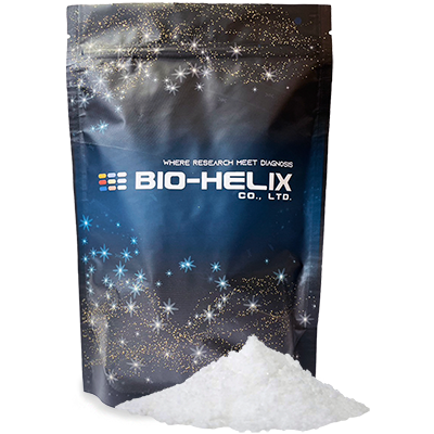
The UltraScence Pico Ultra Western Substrate Powder, as a luminol-based enhanced chemiluminescent substrate, is sensitive and compatible with conducting immunoblots with horseradish peroxidase (HRP) – conjugated secondary antibodies. The low picogram to mid femtogram detection of antigen is enabled by UltraScence Pico Plus-Pico Ultra Western Substrate’s excellent sensitivity and long signal duration. Further, its long chemiluminescent signal duration makes both digital and film-based imaging possible without any loss of the signal. Appropriate primary and secondary antibody dilutions are suggested for attaining optimal signal intensity and duration.
- Ready-to-ship and significantly longer shelf life in the powder form!
- Significant reductions in transportation and storage costs, resulting in less carbon footprint!
- Empower custom production anywhere in the world!
- Significantly more sensitive than leading manufacturers on the market.
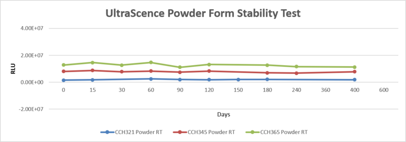
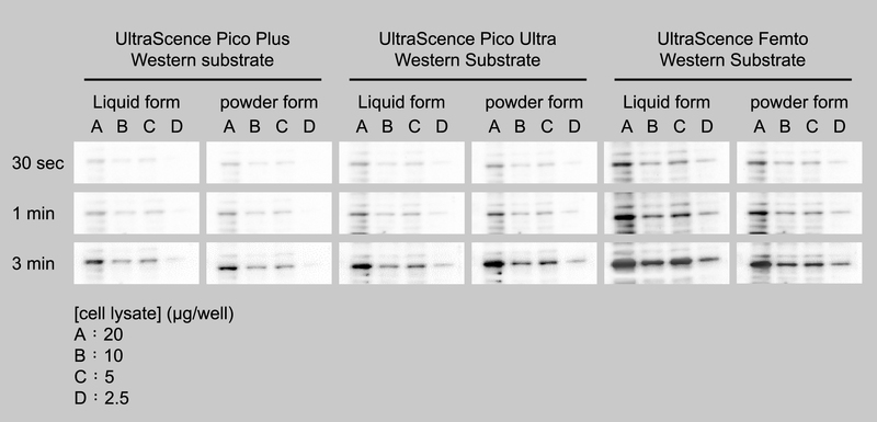
Figure 1. Membranes were probed with Rabbit anti β-actin antibody (GTX109639) diluted at 1:5,000 of and then with Goat anti Rabbit HRP-conjugated secondary antibody (GTX213110-01, 1:10,000) after serial dilution mock RD cell lysates were prepared and applied in electrophoresis and protein transfer. Identical blots were incubated with 400 μL of UltraScence Western substrate. The blots were simultaneously exposed for 30 seconds, 60 seconds and 180 seconds using Chemlux SPX-600 Series digital imaging system.

Figure 2. 24-hour stability test of UltraScence Pico Ultra Western Substrate
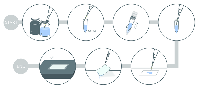
The Luminol and Peroxide solution of UltraScence Pico Ultra were mixed in a 1:1 ratio and having 400 uL treated on each blot after taken out from 4℃ for 1hr, 12 hrs and 24 hrs. The blots were simultaneously exposed for 5, 10, 30, 60, and 180 seconds using Chemlux SPX-600 Series digital imaging system. The EGFP (Enhanced Green Fluorescent Protein) was serially diluted for electrophoresis, after the proteins were transferred to the membrane, they were blocked and probed with GFP tag Rabbit PolyAb (Cat No. 50430-2-AP, 1:10,000), and followed by the Goat Anti-rabbit IgG/HRP secondary antibody (Cat No. SA00001-2, 1:10,000).
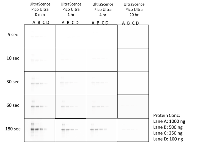
Figure 3. 20-hour signal duration with UltraScence Pico Ultra Western Substrate
The EGFP (Enhanced Green Fluorescent Protein) was serially diluted for electrophoresis, after the proteins were transferred to the membrane, they were blocked and probed with GFP tag Rabbit PolyAb (Cat No. 50430-2-AP, 1:10,000), and followed by the Goat Anti-rabbit IgG/HRP secondary antibody (Cat No. SA00001-2, 1:10,000). Immerse the membrane in a mixed luminol and peroxide solution and check the signal at different times. The blots were simultaneously exposed for 5, 10, 30, 60, and 180 seconds using the Chemlux SPX-600 Series digital imaging system.